Inside a Cannabis Seed: The Part Nobody Photographs


Under the Lens · Episode 7 of 7
A cannabis seed can look like a tiny patterned pebble: a compact oval with an interesting shell. Most photographs stop there. The inside has a different kind of visual appeal, one that only becomes readable when a professionally prepared anatomical image reveals the layers and the folded embryo. Cannabis seed anatomy becomes easier to follow when the fruit wall, seed coat and embryo are identified separately in a prepared section.
Cannabis seed anatomy also comes with a small naming twist. The object commonly called a seed is botanically an achene, a dry fruit surrounding one actual seed. The everyday name is convenient. The botanical distinction explains why the outer shell and the seed coat are not automatically the same structure.
Cannabis seed shell anatomy: the fruit wall
The pericarp is the fruit wall. In cannabis, it encloses a single seed. Both the CFIA biology document and a 2023 AoB Plants microscopy study identify the achene structure.
That is a neat reversal of the ordinary picture. What looks like the outside of the seed in a casual photograph includes a fruit layer. The actual seed has its own covering within that broader object.
You do not need to abandon the everyday word to understand the image. People will keep calling the objects seeds, just as ordinary plant names often compress several anatomical details. The precise terms become useful when a cross-section separates the layers.
A whole-object view and a section therefore answer different questions. The first shows the external shape. The second tells you which visible boundary belongs to which part.
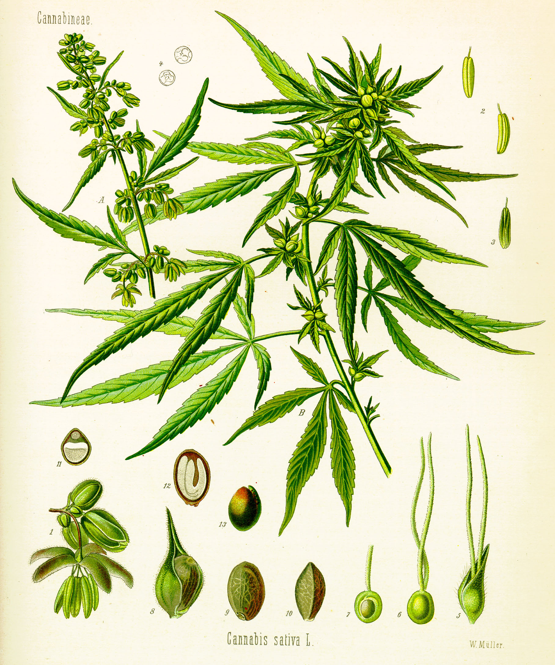
Zoom into the little cutaway drawings
An 1887 botanical plate, in full resolution — The small seed cross-sections around the main plant are easy to miss at first. Open the full-size plate and give them their own close-up.
The embryo is folded into the small space
Kim and colleagues’ 2023 study examined a fibrous hemp cultivar named Chungsam using several microscopy methods. Its anatomical figures identify an embryo with two cotyledons and a radicle, plus endosperm within the seed.
The embryo’s axis is curved into a U-shaped arrangement in the described mature view. That folded layout is the detail that makes the inside surprising. The object does not contain a miniature straight plant standing upright like a toy inside a capsule.
Cotyledons are embryonic seed leaves. The radicle is the embryonic root. Those labels name structures present in the embryo; they are not instructions for planting anything or predictions about an individual specimen.
A properly labeled image lets the viewer follow those parts through the space. The shape becomes more memorable when the caption explains what is curved and what is tucked beside it.
A section is a view through the object
Cross-section and longitudinal section describe different orientations of a cut through a subject. One may reveal how layers sit across its width. Another may show more of the relationship along its length.
That is why two images of the same kind of object can look surprisingly different. A rounded boundary in one view may become an elongated shape in another. The orientation is part of the information, not a minor detail to leave out.
The study’s electron micrographs identify the fruit wall, seed coat and internal structures in prepared views. Their scale bars and labels keep the enlarged shapes tied to real anatomy.
For a visual analogy, think of the difference between a building’s exterior and two floor plans cut at different levels. The analogy helps explain viewpoint. The anatomical image itself supplies the actual shape and labels.
The storage story goes smaller still
Within cotyledon tissue, the study documents oil bodies and protein bodies. Those are cellular structures, at a smaller scale than the whole embryo shown in an anatomical overview.
The shift in scale gives the seed two different hidden interiors. One view reveals the layout of its larger structures. Another reveals material stored inside cells. Calling both images inside the seed is technically understandable, but it hides the enormous difference in what the microscope is resolving.
That is the same reason a whole city map and the interior of one room should not be treated as interchangeable. Both belong to the city; they describe different levels of organization.
You do not need the paper’s detailed biochemical discussion to appreciate the visual distinction. The overview gives the folded body a place. The cell view adds another layer of complexity inside that body.
The labels matter more than a shell-based prediction
An external pattern can be visually interesting without telling the viewer everything about the enclosed tissue. A photograph of the shell is not a viability test, a genetic identification or a guarantee about any future plant.
The anatomical study is also specific to the material its authors examined. It provides real images and an identified cultivar, rather than claiming every object in every casual seed photograph is identical.
Those limits make the close-up easier to trust. The image can be enjoyed for the structures it actually reveals, without turning a pleasing pattern into an unsupported prediction.
There is a useful publication detail here as well. A 2024 correction to the paper updated its title and figure legends, including abbreviation and arrow information. Corrected captions should accompany reused figures so the little labels keep doing their job accurately.
The ordinary oval has an unusually good interior
The shell gives you pattern and shape. The prepared section gives you layers and a folded embryo. The cellular view gives you a still smaller level of organization within that tissue.
Each view changes what the small object feels like. It goes from pebblelike to architectural to cellular, without needing an imaginary cutaway or a claim that someone here performed the experiment.
That is the payoff of a real anatomical photo story. The parts were there before the camera made them readable. A few accurate labels let the reader see why a familiar little oval has far more structure than its outside suggests.
Sources and further reading
- Canadian Food Inspection Agency: biology of Cannabis sativa
- AoB Plants, 2023: cannabis achene and embryo microscopy
- AoB Plants, 2024: corrected title and figure-legend notice
Series cover: original AI-assisted editorial illustration for Trap Snap LA. Photographs carry their own credits.
Frequently asked questions about cannabis seed anatomy
How big is a cannabis seed in millimeters?
CFIA describes cannabis seeds as generally 2–5 millimeters long. That is a botanical range, not a way to identify a cultivar or infer the contents of a particular photographed seed. CFIA cannabis biology document.
What is the perianth around a cannabis seed?
The perianth is floral tissue associated with the outer covering described in CFIA’s seed account. It should not be silently relabeled as the seed coat or the fruit wall, which are different anatomical terms. CFIA cannabis biology document.
What are the layers of the cannabis seed shell?
In the 2023 shell study, the fruit wall had three described zones: exocarp, mesocarp and endocarp. The actual seed coat is another structure; “shell” can obscure that distinction in everyday language. 2023 shell microscopy paper.
What does SEM mean in a cannabis seed anatomy paper?
SEM means scanning electron microscopy. The 2023 shell paper used it to examine fruit-wall and seed-coat structure, so those micrographs should be described as prepared microscope images rather than ordinary exterior photographs. 2023 shell microscopy paper.
Tags: cannabis seed shell layers and microscope images, cannabis seed size and perianth anatomy