
One Flower, Three Levels of Zoom

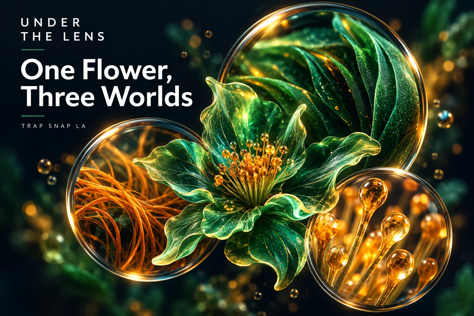
Under the Lens · Episode 1 of 7
One flower can feel like three different subjects depending on how close you get. From a normal viewing distance, it has a shape. In a macro photograph, it has layers. In a microscope image, the surface suddenly has structures that look almost like separate objects.
That is the appeal of macro photography of cannabis flowers: the plant does not need a made-up backstory to be visually strange. Changing the scale changes which details become readable. This is a tour of those levels, using published botanical images as reference points rather than pretending every view came from one photographed specimen.


Level one: the whole shape
An ordinary photograph gives you an outline, a color impression and a sense of how the visible parts sit together. You can see whether a flowering cluster looks narrow, rounded, open or densely layered. You may notice orange-brown strands crossing the surface.
At this distance, the eye tends to combine many small features into a general texture. That is normal. You recognize an object without resolving every part of it, just as you recognize a knitted sweater before you start counting the stitches.
A whole-flower view also gives the close-ups somewhere to live. Without it, an enlarged patch can become an interesting abstract image with no clear relationship to the rest of the subject. The ordinary view tells you where the detail belongs.
Published research often uses this relationship deliberately. Punja and colleagues’ Figure 1 includes whole inflorescence views and closer anatomical photographs. The panels identify different genotypes and specimens, so they are related examples rather than one continuous camera zoom.
Keep zooming
Kandid Kush’s flower and macro portfolio — Move between the wider plant photographs and the extreme details. The same subject suddenly has several completely different looks.
Level two: the surface stops being one texture
A useful close-up lets you separate features the wider picture blends together. Leaflike folds have edges. The longer strands follow their own curves. The fine surface projections can catch the light in small points.
Macro photography has a more specific technical meaning than simply showing a large picture on a screen. Nikon’s macro explanation discusses reproduction ratio: the relationship between the physical subject’s size and the size of its image on the camera sensor. A 1:1 ratio means those physical sizes match.
A giant display therefore does not prove that a photograph was made at high optical magnification. You can enlarge a small portion of an ordinary image until it fills the screen. What matters is whether the capture actually resolved the details you are trying to see.
That is where a good macro image earns its reputation. It can make a rounded gland head or a tiny strand distinguishable instead of merely larger. The difference is between adding screen area and adding visible information.
Level three: the subject becomes a surface landscape
A microscope takes the story beyond what an ordinary flower photograph usually shows. In Figure 2 of the same 2023 paper, the authors identify several trichome forms on sampled cannabis bracts. The views are scanning electron micrographs, with labels and scale bars that explain what is being observed.
A glandular hair with a rounded head can look like a tiny mushroom at this scale. Nearby, a nonglandular hair can look much more pointed. Smaller forms occupy the same surface without sharing the dramatic tall-stalk silhouette.
The analogy is fun, but the labels do the educational work. These are plant-surface structures, not actual miniature mushrooms. Once you know what a head and stalk are, the landscape becomes easier to read.
The sample and imaging method also matter. An electron micrograph is an instrument-produced view of professionally prepared material, rather than an ordinary color photograph made with a phone. Its dramatic appearance comes partly from the way that surface is measured and displayed.
The scale bar is the best part of the caption
A scale bar supplies a physical length. If the picture gets larger on your screen, the bar gets larger with it, preserving the comparison. That is more useful than saying the image is extremely zoomed in.
A micrometer is one thousandth of a millimeter. You do not need to convert every label to enjoy the picture; knowing that relationship is enough to understand why the structures cannot be read clearly in the first view.
The same-sized images on a page can also show completely different physical areas. One may cover most of a flower. Another may cover a small patch of its surface. Equal display size does not mean equal magnification.
That difference is worth pausing over. A tiny patch can fill the frame so convincingly that the original object starts to feel much larger than it really is. The bar brings your sense of size back into the picture.
Zoom reveals structure, not an entire story
At each level, you gain detail and give up some context. The close-up may show a surface beautifully while hiding the whole outline. The microscope may show a gland clearly while showing only a very small sampled area.
No one level has to replace the others. They answer different visual questions: what is the overall shape, what makes the texture, and what structures exist within that texture?
That is the reason to enjoy the whole sequence. The wider image makes the subject familiar. The close view makes it intricate. The micrograph makes it a little alien. It is still the same kind of plant structure, but your brain gets three different ways to appreciate it.
Sources and further reading
- Punja, Sutton and Kim, 2023: cannabis trichome microscopy
- Punja and colleagues: Figure 1, flower and stigma photographs
- Punja and colleagues: Figure 2, identified trichome forms
- Nikon: macro lenses and reproduction ratio
Series cover: original AI-assisted editorial illustration for Trap Snap LA. Photographs carry their own credits.
Questions about cannabis macro photography
What is focus stacking in cannabis macro photography?
Focus stacking combines sharp areas from a series of photographs made at different focus positions. Nikon explains it as a way to increase the depth of field shown in the final composite. Nikon focus-stacking explanation.
Why is only part of a cannabis macro photo sharp?
At close distances, depth of field can be very small. Nikon notes that it may shrink to millimeters in macro work, so a single image can have a narrow sharp region surrounded by blur. Nikon macro-lens explanation.
Does a focus-stacked flower photo mean the plant is fake?
No. A stack can combine real photographs to show more of a real subject in focus. It is still a composite capture method, so an honest caption should distinguish it from a single exposure. Nikon explanation of composite capture.
Do you need a microscope to take macro photos of cannabis?
No. Macro photography can use a camera and a close-focusing lens; microscopy is another imaging method. Nikon describes macro-lens reproduction ratios and close-focus capabilities without requiring a microscope. Nikon macro-lens guide.
Tags: cannabis macro photography focus stacking; why cannabis close up photos have shallow focus.